Human Tooth Structure
Every tooth, whether you are examining a primary incisor or an adult second molar, has the same anatomical structure and components that define the human tooth. The anatomical parts are the anatomical crown, clinical crown, and root(s). The four main components of the tooth structure are enamel, dentin, cementum, and pulp.
Anatomical Structure:
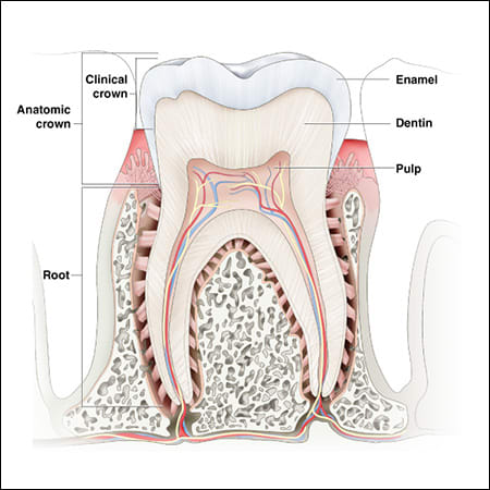
The term anatomical crown is used to define the entire tooth structure that is covered by enamel. This includes areas of the tooth that are covered by the gingival (gum) tissue.
The clinical crown is the area of the anatomical crown that is the visible, exposed structure of the crown. This part of the anatomical crown is not covered by gingival tissue. When you open your mouth and look at your teeth, you are viewing the clinical crown.
The anatomical crown and clinical crown are commonly referred to as the “crown” of a tooth.
The root of the tooth extends into the jawbone and holds the tooth in place with the help of the Periodontal ligament. Depending on the tooth and its primary function, a tooth may have anywhere between one and three roots.
The tooth root contains numerous nerves and blood vessels. The blood vessels supply the Periodontal ligament with the nutrients needed to remain healthy and functioning properly. The nerve bundles send to and receive signals from the pulp to regulate the amount of force used during the act of chewing.
Tooth Components:
Enamel
The crown of the tooth is the outermost layer of the tooth and is covered by a hard semi-translucent layer known as enamel. Enamel is a mixture of minerals (96%), organic tissue, and water, making enamel the hardest substance in the human body. This layer’s sole purpose is to protect the sensitive, delicate inner layers of the tooth where blood vessels and nerve tissue are found.
Dentin
Under the enamel layer of a tooth, is a layer known as dentin. This layer extends from the crown of the tooth to the roots, encasing the innermost layer where the tooth nerve is found. Dentin is stronger than bone but softer than enamel. Like enamel, dentin is also composed of minerals, organic matter, and water.
Dentin supports the structure of enamel, absorbing the natural force a tooth experiences during biting and chewing. This layer is also designed to allow impulses/signals to travel down from the enamel layer to the pulp chamber, which houses the tooth nerve, or up from the roots to the pulp chamber through various small hollow tubes or canals commonly referred to as “root canals”.
Cementum
Cementum is a calcified substance that surrounds the root of the tooth. This layer is softer than dentin and is light yellow in color. Cementum continually forms, replacing older layers with new layers to ensure the attachment of the Periodontal ligament remains.
Periodontal ligament
Each tooth rests in the jaw (alveolar) bone in spaces referred to as tooth sockets. The periodontal ligament (PDL) is a group of soft connective tissue and collagen fiber containing nerves and blood vessels. The primary purpose of the PDL is to connect the tooth socket to the cementum layer of the tooth root, holding the tooth in place. The PDL is also responsible for protecting the tooth and jaw from damage due to the force generated by the act of chewing or external trauma. The flexible nature of the ligament, allows it to function as a “shock absorber”, dispersing the pressure caused by chewing.
Pulp
Beneath the dentin layer is the innermost layer known as the pulp. The Dental pulp is soft and consists of nerves, blood vessels, and connective tissue. This layer is divided into two areas; the pulp chamber located in the crown of the tooth and the pulp canals found in each of the roots. The pulp is responsible for providing nutrients to the tooth and the development of dentin.